
Sports Medicine Resources
STOP Sports Injuries
The National Council of Youth Sports offers safety tips to STOP Sports Injuries. It is a great resource and reminder that our young athletes need appropriate rest and time off from the ever growing world of youth sports.
Visit Site
Shoulder Anatomy
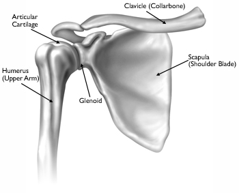 The shoulder is made up of three bones: upper arm bone (humerus), shoulder blade (scapula), and collarbone (clavicle). It is a ball-and-socket joint, meaning the ball, or head, of your upper arm bone fits into a shallow socket in your shoulder blade. This socket is called the glenoid.
The surfaces of the bones where they touch are covered with articular cartilage, a smooth substance that protects the bones and enables them to move easily. A thin, smooth tissue called synovial membrane covers all remaining surfaces inside the shoulder joint. In a healthy shoulder, this membrane makes a small amount of fluid that lubricates the cartilage and eliminates almost any friction in your shoulder.
Together, with the muscles and tendons that surround the shoulder, all of these structures allow the shoulder to rotate through a greater range of motion than any other joint in the body.
Content courtesy of .
Knee Anatomy
 The knee is the largest joint in the body, and one of the most frequently injuried parts of the body.
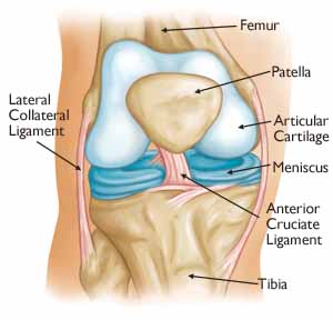
The knee is made up of the lower end of the thighbone (femur), the upper end of the shinbone (tibia), and the kneecap (patella). The ends of these three bones where they touch are covered with articular cartilage, a smooth substance that protects the bones and enables them to move easily.
The menisci are located between the femur and tibia. These C-shaped wedges act as “shock absorbers” that cushion the joint.
Large ligaments hold the femur and tibia together and provide stability. The long thigh muscles give the knee strength.
All remaining surfaces of the knee are covered by a thin lining called the synovial membrane. This membrane releases a fluid that lubricates the cartilage, reducing friction to nearly zero in a healthy knee.
Normally, all of these components work in harmony. But disease or injury can disrupt this harmony, resulting in pain, muscle weakness, and reduced function.
Content provided by .